Certification testing to screen fish for M. cerebralis before export or stocking almost always requires following methodology from the American Fisheries Society-Fish Health Section Blue Book’s USFWS/AFS-FHS Standard Procedures for Aquatic Animal Health Inspections. This test involves performing a pepsin-trypsin digest on cartilage from fresh or frozen samples and then visualizing the spores under a microscope. If myxospores consistent with M. cerebralis are visualized, PCR and histopathology testing can be performed as confirmatory testing.
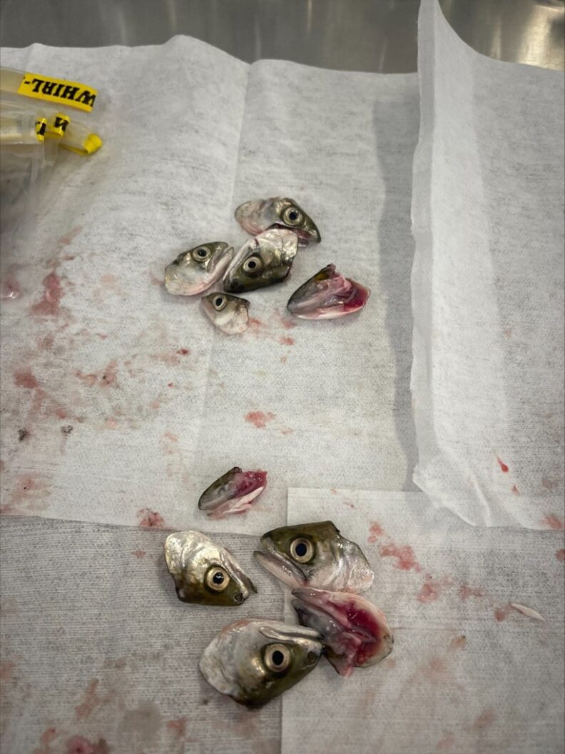
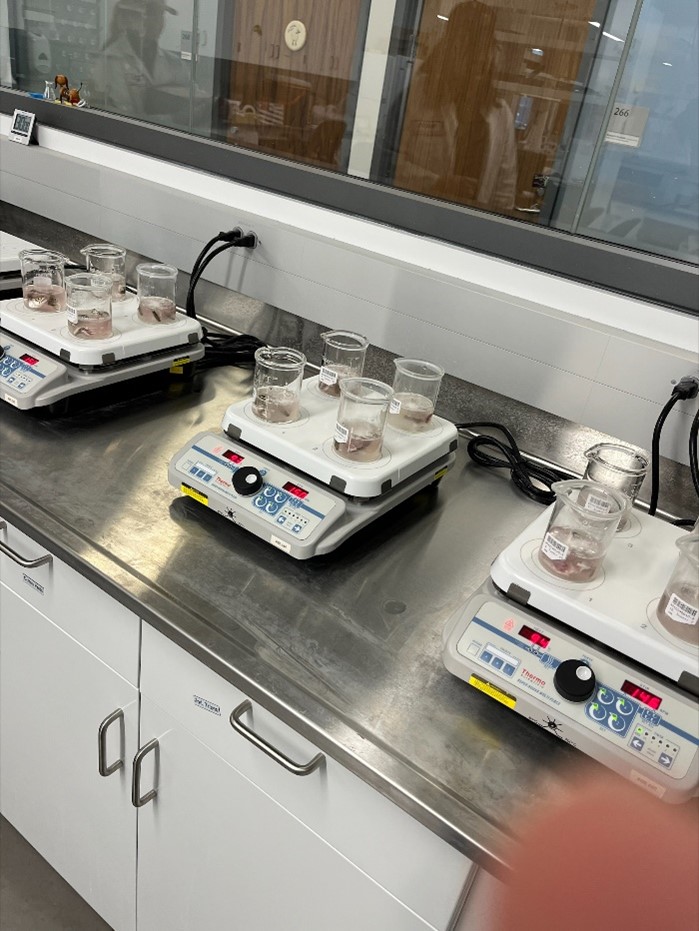
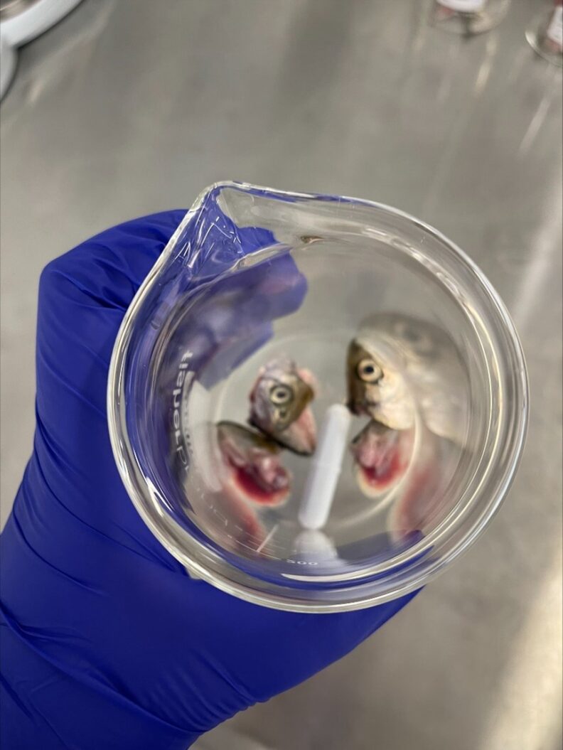
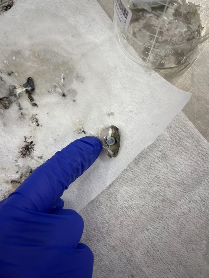
Samples are heated until flesh is soft and eyes are opaque. Then all flesh is removed from the samples and cartilage, bone, gill arches, and opercles are saved for testing.
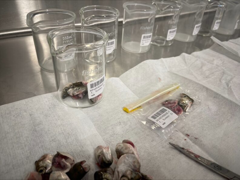
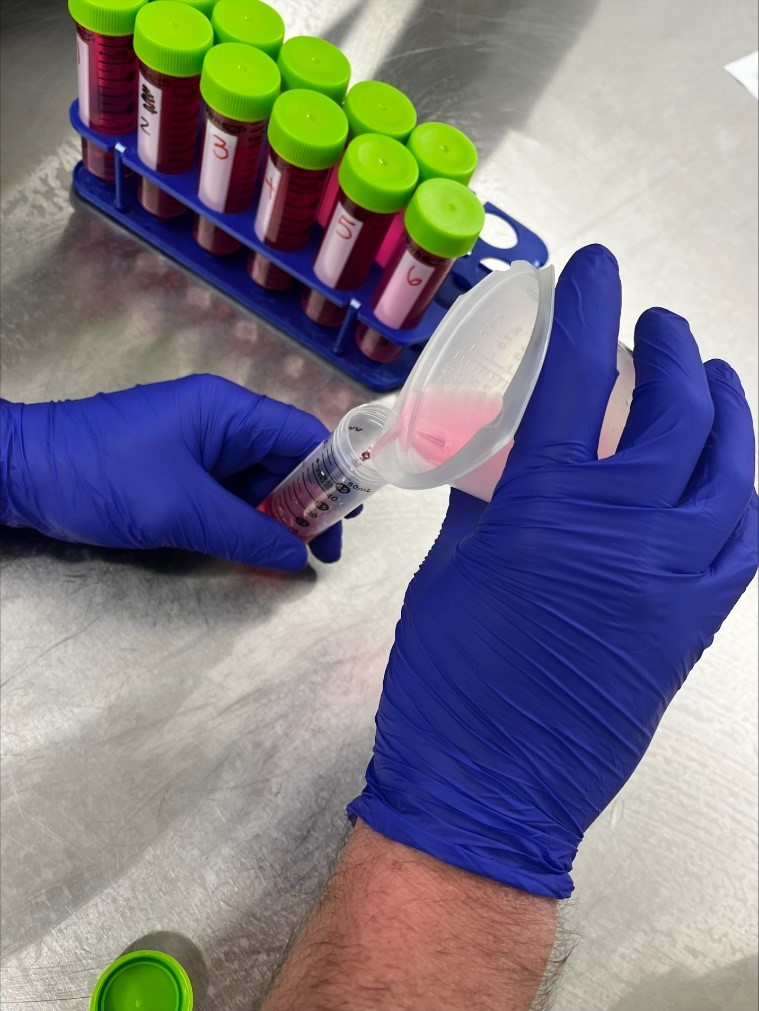
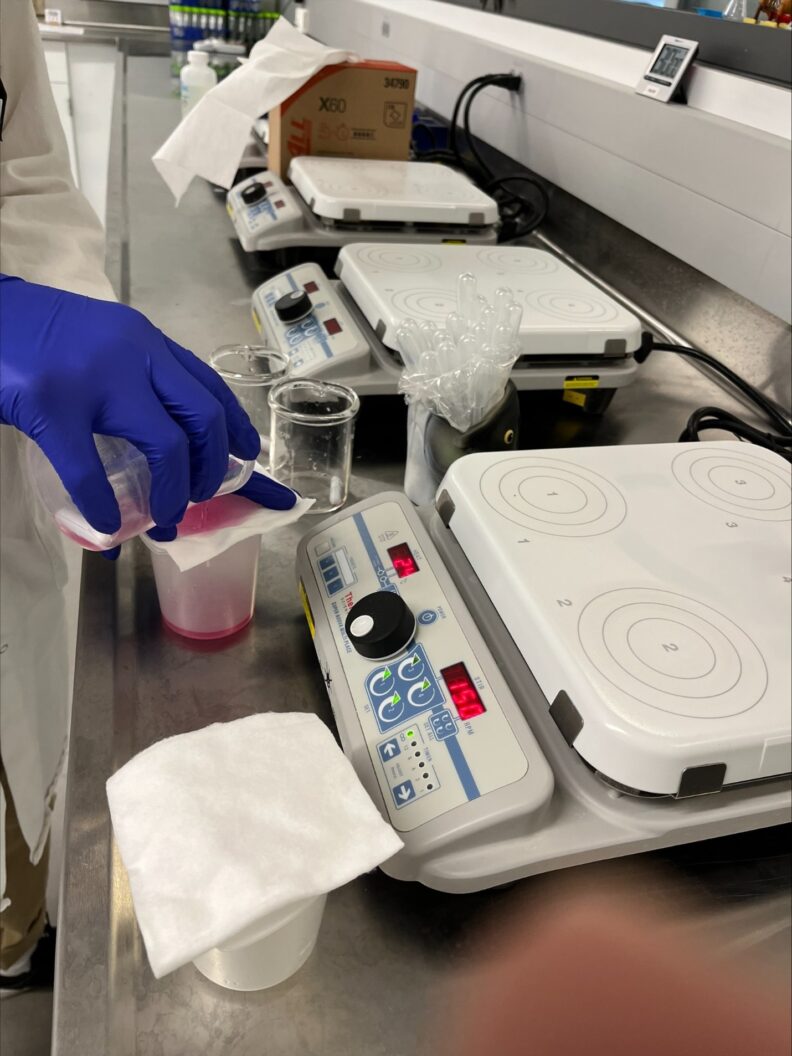
The bone, cartilage, gill arches, and opercles are digested using pepsin and trypsin to small particles to release spores.
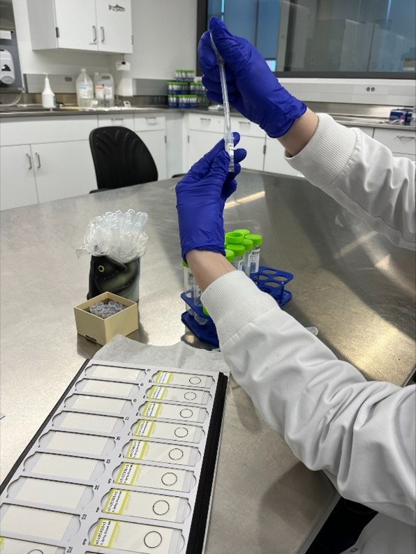
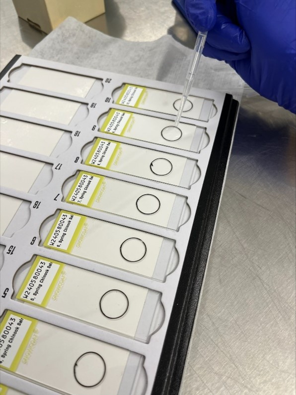
Slides are prepared with pelleted digest material and evaluated microscopically for myxospores.